


Confocal microscopy
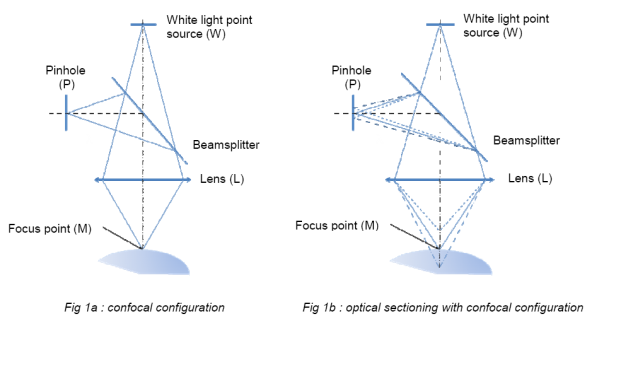
Confocal microscopy is derived from conventional microscopy and uses double spatial filtering. The first filter allows that only one point of the field is illuminated ; the second allows that only the light from the illuminated spot of the object reaches the detector. This configuration is described in Figure 1a, where the point source W is imaged by the lens L in the point M. The reflected light passes through the lens L and is directed to the detector by a divider. The pinhole P plays an essential role in this configuration because it allows only light exclusively from M and filters all, in particular the light from the points above and below M (fig. 1b).
When the scan (x,Y) is added to the confocal configuration, an image of the plane located at a precise distance from the lens L is obtained, without interference from the point outside of the plan. This feature is called “optical sectioning” and is the main benefit of confocal microscopy.
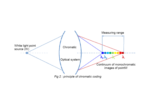
the image of a white light source is a continuous monochromatic dots distributed along the optical axis (fig.2.) Only one of these points is focused on M and thanks to confocal configuration, only the length wave λm back through the spatial filter. If the sample have several transparent layers, each interface between the layers reflects one different wavelength and it's simultaneously detected . The collected spectrum is then decoded to extract the value of the distance from point M.
→ The principle of confocal imaging allows to have excellent spatial resolution whatever the ambient lighting.
confocal spectral interferometry (CSI)
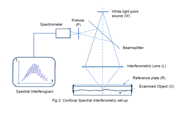
The confocal spectral interferometry is an innovative method to measure the topography and thickness of transparent layers, based on the combination of 2 optical principles: confocal imaging and spectral interferometry.
The Spectral Analysis of interferograms of the White Light (SAWLI) is a method witch consist to analyze the model interference generated by two fronts wave. Usually both fronts wave are generated by reflections on the two transparent sample faces.
One of the alternatives is to place a reference plate sample to create a blade air. Sample thickness (or blade air) is derived from the spectral phase of the interferometric signal, which is calculated with sub-nanometer resolution with an advanced algorithm.
One characteristic of the implanting of the confocal spectral interferometry DUO lies in the fact that the distance between the reference plate and the surface of the sample is fixed during the measurement. The reference plate moves with sample and have the same disturbances, however this don't affect the calculated thickness.